1. What Is Ultrasound?
We encounter three type of sound. Audible, infrasonic and ultrasonic. If we hear any sound that must be in the audible frequency range.
And sound that we cannot hear can be divided into two groups. Ultrasound and Infrasound. We can define these sounds by their frequency range.
• Audible Range: 20 Hz < Audible < 20000 Hz
• Ultrasound: > 20000 Hz
• Infrasound: <20 Hz
What is ultrasound in Medical Industry:
Ultrasound technology or ultrasonography is a method of obtaining image from reflected sound wave.
Ultrasound uses a transducer that simultaneously sends and receives sound waves. The equipment takes photos of soft tissues within the body when a physician glides the probe over a specific gel administered to the testing location.
2. How Many Types Of Ultrasounds Are There?
Ultrasound or ultrasonogram is used in different part of body. We can determine the type by the part is being examined. Here I have discussed about some types you might find helpful.
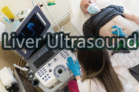
Abdominal Ultrasound
Internal organs such as the liver, gallbladder, spleen, pancreas, kidneys, and bladder can all benefit from an abdominal ultrasound. This can assist in the diagnosis of a range of illnesses as well as the assessment of the damage caused by illness.
Its usage is not limited by that only there are a lot more usages. Here are some more uses of abdominal Ultrasound
1. Needle biopsies, in which syringes are used to extract cells from organs for laboratory testing, are examples of guide operations.
2. Assist a doctor in determining the cause of various stomach problems, such as gallbladder or kidney stones.
3. Assist in determining the source of an abdominal organ's enlargement.
4. Doppler ultrasonography is a type of ultrasound that looks at the significant blood arteries in the body. These photos can assist the doctor in seeing and evaluating:
5. Clots, for example, can obstruct blood flow.
6. Plaque build-up inside the vessel.
7. Malformation that is present at birth.
Pelvic Ultrasound
One of the most well-known types of ultrasound is pelvic ultrasound, which is one of the imaging tests used to evaluate the health of the embryo or fetus throughout pregnancy. These ultrasounds are used to assess the uterus, ovaries, bladder, and prostate gland in addition to maternity medicine.
Some other uses of Pelvic ultrasound are:
1. Pelvic discomfort
2. Bleeding abnormally
3. Problems with menstruation
4. Cysts in the ovaries
5. Fibroids in the uterus
6. Cancers of the ovary and uterus
7. Stones in the kidneys and bladder.
Transabdominal
Patients receiving a transabdominal ultrasound need to have a full urinary bladder. Like other ultrasound procedures, patients lie on their back as a gel is applied to the abdomen. The transducer is then rubbed over the examination are and releases sound waves. This is a fairly straightforward ultrasound exam
Transvaginal Ultrasound
A woman must empty her bladder for a transvaginal ultrasound in the same way she would for a gynecological exam. She also has her feet in stirrups and is lying face up on her back. This test necessitates the use of an ultrasonic transducer.
The transducer is a fraction of the size of a normal Pap speculum. Before inserting the transducer into the vaginal canal, it is covered with a protective cover and lubricating lubricant.
The transducer is only put into the vaginal canal for the first two to three inches. The doctor can move it around to get different viewpoints on the images. Transvaginal pelvic ultrasounds are most commonly used to determine the source of pelvic pain.
Transrectal
During an ultrasound, the transducer must be introduced into the rectum for sound waves to get to the prostate gland. Before insertion, the transducer is protected with a protective cover and lubricated, as with other inserted ultrasonic procedures. The transducer will need to be moved about to collect photos from various angles. The patient is typically examined while lying on their left side with their knees bent up towards their chest.
If a lesion is discovered during the checkup, the doctor may request a biopsy. The radiologist uses ultrasound imaging to guide a needle into the prostate gland and collect a sample of the abnormal tissue during a biopsy. Biopsies guided by ultrasound are less invasive, requiring only a tiny incision.
Obstetric Ultrasound Imaging
Obstetric ultrasonography (OB ultrasound) is a specific use of sound waves for visualizing and determining the status of a pregnant woman and her embryo or fetus. Obstetric ultrasonography should only be used when clinically necessary.
1. To confirm the existence of a live embryo or fetus
2. to figure out how old the pregnancy is
3. Congenital anomalies must be diagnosed.
4. To determine the fetus's position
5. To determine the placenta's position
6. to see if there are any other pregnancies
7. You will be requested to lie backwards or sideways. You'll be requested to expose your lower abdomen as well. It takes roughly 30-45 minutes for an obstetric ultrasound examination.
This process is painless. Pressure from the sonographer guiding the transducer across your abdomen may cause varying degrees of discomfort, especially if you are required to have a full bladder. The sonographer may need to press harder on the embryo or fetus at times to understand the structure better. This discomfort will pass. You may also detest the way the water-soluble gel feels on your abdomen. During transvaginal scanning, there may be some discomfort while the transducer is moved around in the vaginal canal.
Carotid & Abdominal Aorta Ultrasound Imaging
Ultrasound of the carotid artery system is a quick, non-invasive way to detect obstructions in blood flow to the brain in the neck arteries that could result in a stroke or mini-stroke. An abdominal aorta ultrasound is used to check for an aneurysm, which is an abnormal expansion of the aorta caused by atherosclerosis.
The patient is placed on tilting and moving examining table. The area to be inspected is covered with a transparent gel. Because sound waves cannot penetrate air, the gel aids the transducer in making a secure contact and eradicates air pockets between the transducer and the skin.
There are many other types of ultrasound exams, such as those of the liver or vascular system, which are not described here.
However, most ultrasounds take about 30 to 45 minutes to complete, and the patient will receive the results within a few days of the ultrasound. The doctor can use the results to assess the patient's health.