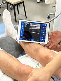
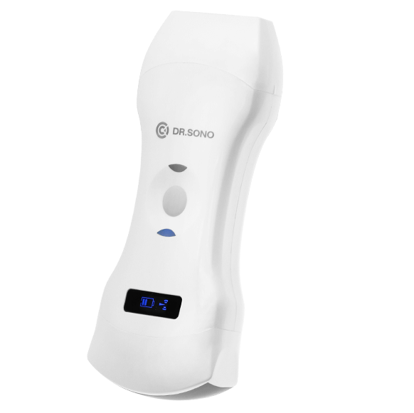
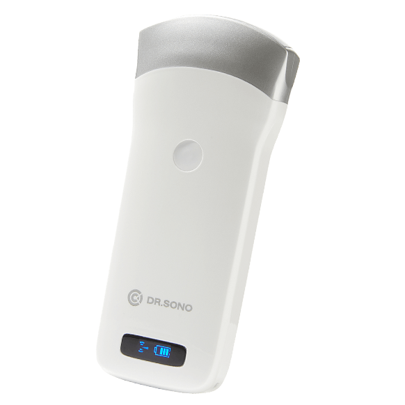
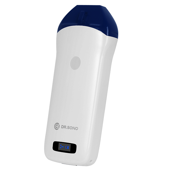
Small Size, Big Features – Perform Whole-Body Scans with 1 Convenient Device
Don’t let its small size fool you. Dr. Sono packs numerous features into its compact design, including a curved probe and a phased array probe. You can choose from 3 scanning modes and just flip the device to go from shallow to deeper imaging.
Linear
Curved
Phased
Avoid Sacrificing Quality for Convenience with Dr. Sono Portable Ultrasound
HD imaging
Enjoy best-in-class HD imaging thanks to this handheld unit’s 192 piezoelectric elements
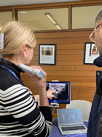
Fit any system
This scanner easily connects to smartphones, tablets, and computers (Windows, Android, iOS)
IPX7
Reduce infection risk with a waterproof scanner that’s easy to clean and disinfect
Wireless
Boost efficiency and flexibility with Internal Wi-Fi and a no-cable ultrasound probe option
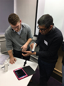
Only 260g
Put this compact, lightweight scanner in your pocket and bring it with you
3 in 1
Select from three scanning modes – convex, linear, and phased – to get the perfect view
Dr. Sono is an FDA Class II registered device that is built using the highest manufacturing standards
Free APP
The Dr. Sono Ultrasound Scanner includes a free app that makes running the device even easier
FREE Shipping
We offer free shipping on US orders. For all other countries, we offer free shipping on orders over $500.
1-Year Warranty
Enjoy peace of mind when you purchase Dr. Sono, it is backed by a 1-year warranty
14 day 100% Money-Back
Dr. Sono also comes with a 14-day, 100% money-back guarantee. If you don’t love it, you can get a refund
Customer Service
Need help or have questions? Simply email us at [email protected] for prompt assistance
FAQ
Can you buy a baby scan machine?
Ultrasound technology has progressed so much that it is possible to purchase a portable ultrasound scanner or even an ultrasound probe that wirelessly connects to your phone and allows you to perform all sorts of ultrasound scans, such as pregnancy scans and abdominal scans and scans for the heart.
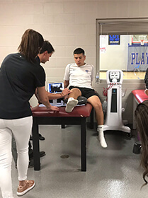
What are the uses of portable ultrasound machines?
Portable ultrasound machines are currently used in cardiac, vascular, radiology, endocrinology, pediatrics, gastroenterology, obstetrics, and gynecology applications.
What is a handheld ultrasound called?
A handheld ultrasound machine is a point-of-care ultrasound that broadcasts ultrasound images to a display tablet or cell phone.
Are Drsono scanners wireless?
Yes, our scanners produce a Wi-Fi signal from a chip located inside the device. This means you can connect to other devices, such as smartphones and tablets, without an Internet connection. You will only need an Internet connection to download the app to operate the scanner. This convenience is why Drsono scanners are popular in both well-populated and remote locations that have limited Internet availability.
Can I do an ultrasound with my phone?
You can use the "wirelessUSG" App on your iPhone or Android phone to examine images from your ultrasound scans at The Ultrasound Clinic. You may effortlessly share your photos with family and friends via social media, instant messaging, and email or simply save them to a safe location.
Hear From Our Customers
We have found the Drsono ultrasound machine to be a great addition to our practice. While the level of imaging may not be as good as a state-of-the-art (and very expensive) ultrasound scanner, it produces excellent images at an inexpensive price and is well suited for rapid diagnosis.
James
Verified customer
Drsono ultrasound scanners are easy to use and very reliable. We have had our scanner for over a year without any technical errors or failures. It hasn't needed repairs at all. Every time we use it, it looks like new
James
Verified customer
Love the product! Super easy to use and delivers great results!
Brian
Verified customer
I believe the Drsono portable ulreasound scanner produces even better image quality than our large cart unit produces. Add to that the fact that it's easier to move around and find the right spot here, and the choice is clear - the Drsono is the best. This scanner is easy to use and easy to clean.
Irma
Verified customer
Drsono POCUS ultrasound scanners are great. They offer everything you could want - from wireless connectivity to small portable size to clear images. I highly recommend these devices
Dorothy
Verified customer
I recently ordered the wireless probe from DRSONO. I had no problems cleaning the transducer. There are no wires so it is very easy to move. Once a drop of water fell on the transducer, no damage occurred. The image quality is very high and user friendly.
Randy
Verified customer
Join Our Mailing List & Save!
Enter your email address below to receive exclusive promotions and discounts along with additional product information and tips