1. What Is Ultrasound?
Many people associate the phrase "ultrasound" with a pregnant lady at her doctor's office getting a glimpse at the baby growing within her womb – possibly even deciding whether to paint the nursery pink or blue. While prenatal imaging is one of the most prominent applications for ultrasounds, this diagnostic tool has numerous other uses.
Diagnostic ultrasonography, often known as sonography or diagnostic medical sonography, is a type of imaging that employs sound waves to create pictures of structures within your body. The photos can give helpful information for diagnosing and treating various illnesses and ailments.
Most ultrasound tests are performed using an ultrasound device outside your body, while some need the placement of a tiny device within your body.
An ultrasound is performed by passing a device known as a transducer or probe over a part of your body or within a bodily orifice. The provider applies a tiny gel coating to your skin, allowing the ultrasound waves from the transducer to pass through the gel and into your body.
The probe turns electrical current into high-frequency sound waves and transmits them into your body's tissue. The sound waves are inaudible to you.
Sound waves bounce off structures within your body and return to the probe, which converts them into electrical impulses. A computer then converts the pattern of electrical impulses into real-time pictures or films projected on a nearby computer screen.
2. History Of Ultrasound
Ultrasound in medicine was first used in several settings worldwide during and soon after World War II. The oldest known work on medical ultrasonics is Dr. Karl Theodore Dussik's 1942 study on transmission ultrasound studies of the brain in Austria.
Although many individuals in the United States, Japan, and Europe have been acknowledged as pioneers, Professor Ian Donald and his colleagues at Glasgow in the mid-1950s made substantial contributions to developing practical methodologies and applications.
This led to the widespread use of ultrasonography in medical practice during the next several decades.
What are the different types of ultrasounds and their purposes?
Ultrasound imaging is divided into three major groups, which are as follows:
• Ultrasound during pregnancy.
• Ultrasound for diagnosis.
• Procedures are guided using ultrasound.
Ultrasound during pregnancy
During pregnancy, healthcare practitioners frequently utilize ultrasonography (also known as prenatal or obstetric ultrasound) to monitor mother and the baby.
Prenatal ultrasound is used by providers to:
Confirm pregnancy.
Check to find out whether you're expecting more than one child.
Calculate the length of your pregnancy and the gestational age of your unborn child.
Examine your unborn child's fetal development and posture.
View your unborn baby's activity and heart rate.
Examine your unborn baby's brain, spinal cord, heart, and other organs for congenital disorders (birth abnormalities).
Examine the amniotic fluid level.
At 20 weeks pregnant, most doctors recommend getting an ultrasound. This test monitors the growth and development of your unborn child during pregnancy. This ultrasound may reveal your baby's biological sex as well. Tell your technician if you want to know the sex or not.
Your provider may request further scans to address any questions or concerns, such as the possibility of congenital diseases.
3. Ultrasound For Diagnosis
Diagnostic ultrasounds are used by providers to inspect inside portions of your body to determine whether something is incorrect or not operating properly. They can assist your provider in learning more about what's causing a variety of symptoms, such as unexplained discomfort, masses (lumps), or an abnormal blood test.
The transducer (probe) for most diagnostic ultrasonography tests is placed on your skin by the technician. In other situations, the probe may need to be inserted into your body, such as your vagina or rectum.
The sort of diagnostic ultrasonography you receive is determined by the specifics of your situation.
4. Diagnostic Ultrasounds Include The Following
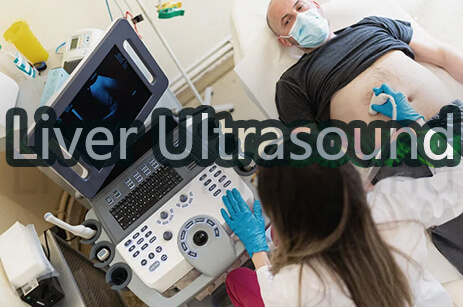
Ultrasound of the abdomen: An ultrasound probe glides across the skin of your stomach (belly). Many causes of stomach discomfort can be diagnosed using abdominal ultrasonography.
Kidney (renal) ultrasound: A kidney ultrasound is used by providers to check the size, position, and form of your kidneys and associated organs such as your ureters and bladder. Cysts, tumors, blockages, and infections in or around your kidneys can be detected using ultrasound.
Breast ultrasound: A breast ultrasound is a noninvasive diagnostic used to detect tumors and cysts in the breast. Following abnormal mammography, your physician may advise you to get an ultrasound.
Doppler Ultrasound: Doppler ultrasonography is a type of ultrasound that measures the movement of elements in your body, such as blood. It enables your physician to observe and analyze blood flow through your body's arteries and veins. Doppler ultrasound is frequently utilized in diagnostic ultrasound studies or vascular ultrasounds.
Pelvic ultrasound: A pelvic ultrasound examines the organs in your pelvic region, located between your lower abdomen (belly) and your legs. Your bladder, prostate, rectum, ovaries, uterus, and vagina are all pelvic organs.
Transvaginal ultrasound: Your physician inserts a probe into your vaginal canal. It depicts reproductive structures like your uterus and ovaries. Because it assesses systems inside your pelvis, a transvaginal ultrasound is also known as a pelvic ultrasound (hip bones).
Thyroid ultrasound: Ultrasound is used by providers to evaluate your thyroid, a butterfly-shaped endocrine gland in your neck. Providers can assess the size of your thyroid gland and look for nodules or abnormalities inside the gland.
Transrectal ultrasonography: Your clinician inserts an ultrasound probe transducer into your rectum. It examines your rectum or other surrounding tissues, such as the prostate, in men who are born male.
5. The Advantages Of Getting An Ultrasound
Many patients and clinicians prefer ultrasonic diagnostic imaging tests to other diagnostic imaging procedures. To view tissues in the body, ultrasound methods use sound waves and their echoes.
Because of its safety and ease, an ultrasonic scan has numerous unique benefits over other types of scans. The following are some of the advantages of ultrasound:
(1)Visualization Of Soft Tissue
Soft tissues, such as organs and muscles, are exceptionally well seen with ultrasound technology. When sound waves strike tissues of varying densities, an echo reflects and is recorded using ultrasound equipment.
The soft tissue determines the echo's properties, such as intensity and timing. Tumors and other abnormalities are visible on ultrasonic scans because their density differs from surrounding tissue.
Some imaging modalities, such as X-rays, for studying soft tissue, are less trustworthy. These tests are more effective on hard tissues like bone.
(2) Non-Invasive Methodology
The ultrasound technician usually only needs to attach the probe to the areas that require vision, such as a pregnant woman's abdomen or the neck of someone having their thyroid gland examined. Other times, the radiologist may need to insert the probe into bodily cavities to obtain pictures of specific organs, although this does not need to break the skin or cause an impact on the body. A transvaginal ultrasound, for example, is required to obtain good images of the uterus and ovaries.
Ultrasound is sometimes used to guide real-time surgeries, such as putting a probe within the body. Even in these cases, the ultrasound process does not require the skin to be broken.
(3) Accessibility And Quickness
Ultrasound sessions are typically brief, lasting only a few minutes. Even the most extensive ultrasound scans take no more than an hour. As a result, ultrasound tests are handy for people with hectic schedules.
In comparison to other imaging services, ultrasound is also quite affordable. The only component required for ultrasonic imaging is a water-based gel that helps the probe's signals penetrate the skin and organs. The cheap expenses equate to a smaller medical bill, which is another reason doctors prescribe ultrasound testing first if appropriate.
(4) Safety
Ultrasound imaging produces pictures purely via the use of high-frequency sound waves. Unlike other imaging tests, ultrasounds do not use radiation. Thus, an ultrasound scan cannot cause radiation-related health concerns.
Furthermore, several imaging modalities require using compounds known as contrast agents. These substances are necessary to highlight specific body concerns during diagnostic imaging. Contrast agents are always administered orally or by injection, allowing these compounds to circulate throughout the body. Some persons are allergic to various chemicals, and ultrasound can function without them, making the scanning procedure safer.
Even after decades of usage, there are no reported health hazards or adverse effects from exposure to ultrasound's high-frequency sound waves. Because of this high level of safety, many healthcare practitioners advocate ultrasound testing as a first-line imaging modality.
(5) Uses Of Ultrasound Scan
Ultrasound imaging is used in medicine for various purposes, including confirming and dating a pregnancy, diagnosing certain illnesses, and directing doctors through precise medical operations.
Pregnancy. During pregnancy, ultrasound pictures can be used for a variety of purposes. They can be used to establish due dates, indicate the presence of twins or other multiples, and rule out early ectopic pregnancies. They are also useful screening techniques for detecting possible difficulties, such as birth abnormalities, placental complications, and breech placement. Midway through the pregnancy, many pregnant parents look forward to knowing the sex of their baby via ultrasound. Later in pregnancy, ultrasounds can even be used to determine the size of a baby right before delivery. Even after decades of usage, there are no reported health hazards or adverse effects from exposure to ultrasound's high-frequency sound waves. Because of this high level of safety, many healthcare practitioners advocate ultrasound testing as a first-line imaging modality.
Disease Detection: Doctors use ultrasound imaging to diagnose various disorders affecting the body's organs and soft tissues, including the heart and blood arteries, liver, gallbladder, spleen, pancreas, kidneys, bladder, uterus, ovaries, eyes, thyroid, and testicles. However, ultrasounds have certain diagnostic limitations; sound waves may not pass effectively through solid bone or regions of the body that may contain air or gas, such as the colon.
During medical operations, use. Ultrasound imaging can assist doctors in performing procedures such as needle biopsies, which involve the removal of tissue from a very exact region inside the body for testing in a lab.
(6) Ultrasound For Soft Tissues
Ultrasound creates pictures by using high-frequency sound pulses. A transducer is a tiny handheld device that sends and receives sound waves. The visuals are created by using the returning sound waves. New ultrasound technology allows us to examine soft tissue anomalies surrounding joints, tendons, and muscles in incredible detail. In skilled hands, it is an exact method that may be used to identify a wide range of soft tissue disorders.
Musculoskeletal ultrasound for traumatic, inflammatory, and degenerative conditions
Musculoskeletal ultrasonography is most commonly used to assess the traumatic, inflammatory, and degenerative states of musculoskeletal structures such as joints, tendons, ligaments, and muscles. This method is widely used to identify tendon rips or inflammation, particularly in the shoulder rotator cuff and the ankle's Achilles tendon.
Muscle and ligament injuries or sprains are also routinely detected with this approach. This method may readily detect soft tissue masses with diameters smaller than 5 cm; moreover, the system can see fluid buildup inside soft tissue, joint effusions, synovial membrane inflammation, and peripheral nerve lesions.
It cannot identify masses that are too huge or too deep inside the tissue nor discover causes of poorly localized pain or other nonspecific symptoms.
Due to high-quality imaging, musculoskeletal ultrasonography has become a popular method for identifying benign and malignant soft tissue tumors, hernias, and ganglion cysts. The approach can also detect pathological alterations linked with illnesses like rheumatoid arthritis and carpal tunnel syndrome.
Musculoskeletal ultrasonography is often utilized in pediatric healthcare to diagnose hip dislocation, neck muscle abnormalities, soft tissue masses, and fluid buildup in the hip joint.
Ultrasound can assist diagnose active inflammations by detecting enhanced blood flow in soft tissue using color flow imaging or color Doppler imaging.
Musculoskeletal ultrasonography is used therapeutically to steer the needle during steroid injections into specific joints and nearby soft tissue to relieve joint pain and aspirate fluids from particular target locations. Furthermore, this approach makes it simple to visualize the post-treatment healing phase.
(7) Ultrasound Scanner At Drsono
DrSono is one of the best online shops that will provide you with the best portable ultrasound scanner. You can find different types of ultrasound scanners for different purposes. So contact DrSono now for the best ultrasound experience.