Taking an ultrasound for diagnostic or other medical purposes has become more common, and with it, the use of related diagnostic terms among non-medics. Sometimes, however, terms used as synonyms in the medical context can spin the heads of patients and the public.
Our case in point? Ultrasound vs Sonogram.
So, are ultrasound and sonogram the same or different?
There’s a slim technical difference between the words ultrasound and sonogram. But the two are aspects of the same procedure. We’ve laid out the differences and comparisons in this article. Let’s take each term at a time, starting with ultrasound.
What is an Ultrasound?
The origin of ultrasound is in two adjectives: “ultra” meaning beyond the ordinary or the limit, and “sonic” which points to the nature of sound speed.
Put together, these two words form a compound adjective describing sound that has a frequency beyond human hearing capacity (>20 kHz).(1)
The medical world borrows the original meaning of ultrasound to describe medical imaging that uses high-frequency sound waves to create images of internal body organs and structures.
So, how do sound waves turn into images? That’s what the next section is about.
How Does an Ultrasound Work?
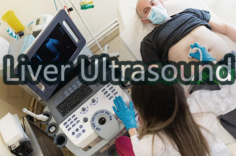
The ultrasound medical procedure relies on imaging equipment known as an ultrasound machine.
This machine has four main parts: a computer processor, a transducer or probe, a monitor or screen, and a printer.
During the ultrasound process, an ultrasound technician passes the transducer on the skin over the area under examination. The transducer sends high-frequency sound waves to the organ or tissue in focus.
Once these waves hit the targeted organ or tissue, they are reflected back to the transducer as electric signals. The transducer then sends these signals to the computer processor, which turns them into the images displayed on the monitor.
The ultrasound tech can then create hard copies of the images using the printer. S/he can also interpret them from the screen and then send a report to the requesting doctor for diagnostic purposes
What is an Ultrasound Used for?
Ultrasounds have different purposes, including the following:
Medical Diagnosis
In diagnostic ultrasound, technicians use the ultrasound procedure to create images of body organs that doctors can use as a diagnostic aid.
Ultrasounds are non-invasive imaging procedures, making them safer than procedures that use ionizing radiation, such as X-rays and CT scans. Experts associate these radiation imaging procedures with a risk for cancer and allergic reactions.(2)
Examples of ultrasound procedures that produce images for medical diagnosis are abdominal, pelvic, muscle, and heart ultrasound, also known as echocardiogram.
Fetal Development Monitoring
Fetal development monitoring ultrasounds create images of a fetus in a mother’s womb. OB/GYN doctors use these images to tell mothers how far along they are in their pregnancy and how their baby is doing in the womb. The ultrasound can also show the baby’s sex by the end of the first trimester or the beginning of the second.
Medics and mothers prefer obstetric ultrasounds because they are free of exposure to radiation and, therefore, safe for both mother and baby.(3)
Therapeutic Ultrasound
Ultrasound technicians direct therapeutic ultrasound processes to body tissues to alter or destroy them. Ultrasound processes used for therapeutic purposes can:
- Help deliver drugs to precise body locations.
- Push or move body tissues.
- Heat body tissues.
- Dissolve blood clots.(4)
All these therapeutic ultrasound functions are non-invasive and do not involve any surgical operations, which saves patients the horror of post-therapeutic scars.
But what about a sonogram? Is it the same as an ultrasound?
What is a Sonogram
A sonogram is an image of internal body organs resulting from the ultrasound procedure, also called sonography.
The two words that make the term sonography point back to the ultrasound procedure explained above: “sono” (relating to sound) and “graph” (recording/writing process).
So, sonography is the recording of sound or sound waves to create an image or sonogram.
What is a Sonogram Used For?
When an ultrasound technician or sonographer produces a sonogram, they send it to the requesting doctor for diagnostic and treatment purposes. The doctor has to read the sonogram.
Interpreting a sonogram is possible because once the sound waves from the transducer reach body structures or tissues, the tissues reflect them back to the transducer at different intensities depending on their echogenicity or ability to reflect.(5)
The reflection intensity of soundwaves makes them appear in different color shades on the ultrasound machine screen:
- Bones and solid masses return sound waves in high intensity and appear in white.
- Less dense body tissues reflect sound waves in lesser intensity and are decoded in a gray shade.
- Liquid body fluids absorb sound waves and don’t reflect them back. So, they appear black on an ultrasound image.
Using these colors, doctors diagnose abnormal cells and body structures in their patients or tell the development progress of fetuses in their mothers’ wombs.
In Summary
What’s the difference between a sonogram and an ultrasound?
An ultrasound is a non-invasive procedure in medical imaging that uses high-frequency sound waves. A sonogram is the image produced through an ultrasound procedure.
As such, ultrasound and sonogram are aspects of the same procedure.
So, when explaining ultrasound vs sonogram pregnancy tests, we would refer to the imaging procedure as an ultrasound to monitor fetal growth and a sonogram as the resulting image of the fetus.
Ultrasound images or sonograms aid diagnosis and treatment and help monitor fetal development. The ultrasound also assists in direct therapeutic procedures.
What is the Difference Between Sonography and Ultrasound Technician?
Sonography is the diagnostic procedure that uses high-frequency sound waves produced by ultrasound machines to produce images of internal body organs known as sonograms. An ultrasound technician is a professional who operates the ultrasound machine used in sonography to produce sonograms.