Ultrasound and X-ray are two revolutionary machines for medical examinations. To detect diseases, doctor’s sometimes need to sneak inside the patient's body to get a good look at what's happening.
1. What Is An Ultrasound Scanner?
Ultrasound technology is a fantastic diagnostic tool for seeing live pictures of the functional structures of the body, particularly the structures of joints. Ultrasound imaging (sonography) creates a live video feed image of the inside of the body by using high-frequency sound waves. Ultrasound is the technology, or "eyes," that allows doctors to obtain a closer look to provide an accurate diagnosis.
Because ultrasound pictures are collected in real-time, they may also reveal the movement of the body's interior organs and blood moving via blood veins. Unlike X-ray imaging, ultrasonic imaging does not involve any radiation exposure.
2. How Does Ultrasonography Work?
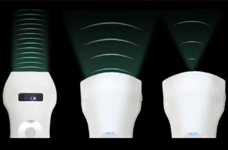
Ultrasound imaging uses a tiny transducer (probe) to send sound waves into the body and record the waves that return. Sound waves go through the investigated region until they reach a tissue border, such as the boundary between the fluid and soft tissue or soft tissue and bone. Some sound waves have reflected the probe at these limits. In contrast, others travel further until they hit another barrier and are reflected. Because the waves' velocity, direction, and distance traveled vary based on the barrier they encounter, a computer may interpret this data as a two-dimensional picture on a screen.
The waves' reflection determines the quality of ultrasound pictures of bodily structures. The information required to construct an image is provided by the strength or amplitude of the sound signal and the time it takes for the wave to pass through the body. These photos assist the doctor in more closely examining something and making an accurate diagnosis.
3. What Is X-Ray?
Medical radiology began in 1895 when Wilhelm Röntgen developed what is now known as the X-ray. Röntgen was investigating how electrical rays could flow from an induction coil through a glass tube at the time. The tube was covered in dark paper in an entirely dark room, and he saw that the rays emitted by the glass tube lighted a screen coated in fluorescent material.
This was the beginning of Röntgen's realization that these rays might be employed to pass through other things. Instead of a screen, he utilized an image of his hand on a photographic plate, which was projected translucently, providing an internal representation of his hand's anatomy. This discovery demonstrated that the inside portions of our bodies might be examined without invasive and hazardous surgery. The advent of the X-ray aided in the modernization of medicine, growing into what we now call the digital X-ray.
It was soon realized that repeated X-ray exposure may be detrimental, although specific precautions are now taken to safeguard the patient and the doctor and avoid problems. Digital radiography provides various advantages over traditional film/screen X-rays today, including lower radiation, higher picture quality for accurate diagnosis, and faster results.
4. How Does X-Ray Work?
An x-ray machine is simply a form of camera with particular capabilities. Instead of using visible light to expose the film and generate a picture, x-rays use radio waves to expose the film and make an image. Bone tissue absorbs many X-rays because it is high in calcium, but soft tissues like fat and skin do not. Areas with less thick tissue seem darker in an X-ray picture, whereas regions with denser tissue appear brighter. This is why X-rays show bones and metal objects as white.
5. What Is The Difference Between X-Ray And Ultrasound?
We can discuss the differences between ultrasound and x-ray by categorizing them by some criteria.
The waves' nature:
An X-ray is a transverse wave. The propagation does not require a material medium.
A longitudinal wave is what ultrasound is. The propagation requires a material.
Frequencies:
The frequency of X–rays ranges from 1016 Hz to 1020 Hz.
Ultrasound frequencies range from 2 to 12 MHz
Applications:
Some X-ray uses include X-ray fluorescence (non-destructive elemental analysis), radiography in medicine, X-ray lithography, X-ray treatment, X-ray
crystallography, and so on.
Ultrasound waves are utilized in various applications, including ultrasound imaging, sonar devices, non-destructive testing, acoustic microscopes, and ultrasound cleaning.
Ionizing Capability:
X-rays can ionize atoms. Atoms cannot be ionized by ultrasound.
Risk:
Because X-rays are high-energy waves, they can interact with DNA and cells. The possibility of X-rays causing cancer is a danger.
Mechanical acoustic waves are what ultrasound waves are. As a result, they pose no danger.
Details Detection:
Both ultrasound and X-rays are critical digital imaging technologies used in veterinary medicine. An X-ray focuses on the bones, lungs, and gas-filled organs, whereas an ultrasound concentrates on the organs' interior intricacies.
When diagnosing the heart, soft tissues, fluid buildup, and so on, ultrasound is far more accurate and beneficial.
Safety:
As previously stated, excessive X-ray radiation exposure harms both the animal and the person holding it. X-rays should not be performed on pregnant horses since unborn offspring are vulnerable to these types of radiation.
Ultrasound imaging of a horse, on the other hand, is harmless since it employs sound waves and is not hazardous. As previously stated, ultrasonography radiation is a sound wave and is safe for pregnant equines.
Abnormal Fluid Accumulation Detection:
Soft tissues and fluids have the same radiological density. So, radiography cannot distinguish between them. However, ultrasound radiation can easily separate liquids from soft structures such as the spleen and liver.
Although the presence of fluids in the body reduces the utility of X-ray pictures, ultrasonography takes advantage of it.
Fluid in the body permits ultrasonic radiation to move quickly, allowing veterinarians to check deep-seated organs. This benefit enables ultrasonic imaging to identify probable causes of fluid buildups, such as pericardial effusion and hemo abdomen.
Heart Disease Detection:
Cardiovascular studies, or echocardiography, are one of the most prevalent uses of ultrasound imaging. Vets use echocardiography to assess the blood flow, cardiac chambers, and heart and valve functioning. Radiography can determine whether or not a heart is enlarged, but it cannot determine why.
Ultrasound radiation reports, on the other hand, give specific information about the heart and allow veterinarians to provide life-saving therapy on time. The Doppler effect in ultrasound radiation helps detect cardiac problems in horses.
6.Conclusion
Ultimately, we can say X-ray and ultrasound are both important inventions for the medical world. It would be best if you choose according to the doctors' advice.
Stay healthy, Stay Safe.