Ultrasound and CT scans are two tests used to screen people for cancer and other abnormalities. Although they are both imaging instruments, there are considerable distinctions between the two tests and what they are capable of detecting.
If you are confused about these imaging technologies, the following article is just for you.
Let us first check out what is ultrasound scanning and CT scanning.
1. What Is Ultrasound?
An ultrasonic scan creates pictures of the interior of the body by using high-frequency sound waves. It is safe to use when pregnant.
Sonography, or ultrasound scans, are safe because they create pictures using sound echoes and waves rather than radiation.
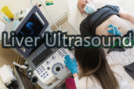
Ultrasound scans are mainly applied to assess fetal development and can discover liver, heart, kidneys, or abdomen issues. They may also assist with some types of biopsies.
The resulting picture is known as a sonogram.
Ultrasounds have several restrictions regarding the structures they can detect, often restricted to internal organs. Ultrasounds are rarely requested for imaging of bone structures.
2. History Of Ultrasound Scanning
The use of ultrasound in medicine was introduced during and immediately after World War II in a variety of locations across the world. Dr. Karl Theodore Dussik's study on transmission ultrasound research of the brain in Austria in 1942 is the earliest documented work on medical ultrasonics.
Although various workers in the United States, Japan, and Europe have been credited as pioneers, Professor Ian Donald and his colleagues at Glasgow in the mid-1950s contributed significantly to the development of practical techniques and applications.
This resulted in the widespread use of ultrasonography in medical practice throughout the following decades.
3.How Does Ultrasound Work
Ultrasound imaging works by sending sound waves into the body and recording the waves that return. Sound waves travel through the researched region until they encounter a tissue barrier, such as the boundary between the fluid and soft tissue or soft tissue and bone. At these limitations, some sound waves have reflected the probe. Others, on the other hand, travel until they meet another barrier and are reflected. Because the waves' velocity, direction, and distance traveled vary depending on the wall they hit, a computer may interpret this data as a two-dimensional image on a screen.
The reflection of the waves determines the quality of ultrasound images of bodily structures. The power or amplitude of the sound signal and the time it takes for the tide to transit through the body offer the information needed to generate a picture. These images let the doctor examine things more closely and diagnose accurately.
4. What Is A CT Scan?
Medical practitioners utilize computed tomography to analyze structures within your body, generally known as a CT scan. A CT scan creates pictures of a cross-section of your body using X-rays and computers. It captures photos of your bones, muscles, organs, and blood arteries, allowing healthcare specialists to observe your body in incredible detail.
Traditional X-ray machines employ a fixed tube to direct X-rays to a specific location. As X-rays flow through the body, various tissues absorb varied quantities of them. Higher density tissues provide a whiter picture than lower density tissues against the film's black backdrop. X-rays generate two-dimensional images. CT scans use a doughnut-shaped tube that circulates the X-ray around you 360 degrees.
5. History of CT Scan
In 1972, British engineer Godfrey Hounsfield of EMI Laboratories developed the first commercially accessible CT scanner. Dr. Allan Cormack, a physicist, co-invented the technique. Both researchers were later given the Nobel Prize in Physiology and Medicine in 1979. By 1981, Hounsfield had been knighted and was known as Sir Godfrey Hounsfield.
However, Johann Radon's mathematical theory, known as the "Radon transform," published in 1917, gave birth to the technology. The "Algebraic Reconstruction Technique," developed by Polish mathematician Stefan Kaczmarz in 1937, is another mathematical innovation on which Hounsfield built. Hounsfield used both theories to make one of the most significant advances in medical history.
6. How Does CT Scan Work?
Unlike a traditional x-ray, a CT scanner employs a motorized x-ray source that spins around the circular entrance of a donut-shaped frame called a gantry. The patient rests on a bed that gently travels across the gantry as the x-ray tube rotates around the patient, sending narrow beams of x-rays through the body during a CT scan. CT scanners employ sophisticated digital x-ray detectors instead of film, which are immediately opposite the x-ray source. The sensors take the x-rays as they leave the patient and send them to a computer.
The CT computer employs advanced mathematical procedures to produce a two-dimensional picture slice of the patient every time the x-ray source completes one full revolution. The thickness of the tissue in each imaging slice varies depending on the CT equipment used, although it typically ranges between 1 and 10 millimeters. When a whole piece is finished, the picture is saved, and the motorized bed is slowly dragged ahead into the gantry. The x-ray scanning procedure is then repeated to generate another picture slice. This technique is repeated until the necessary number of pieces is obtained.
7. Difference Between CT And Ultrasound Scan:
Ultrasound Scans and CT scans both are methods to image your internal body. From their procedure of work, I have given above will enlighten you about it. But their difference does not end here. There are different categories on which we can differentiate them. I have discussed those parameters below.
Exposure to Radiation
Sonography, often known as ultrasound, creates a picture of inside structures by employing high-frequency sound waves. There is no radiation exposure when using ultrasonography. However, there is some radiation exposure with a CT scan, around the same that an average person receives from ambient radiation in 3 to 5 years. CT scans are not suggested for pregnant women or children due to the radiation risk they involve.
Image Recognition
Ultrasounds have several restrictions in terms of the structures they can detect, which are often restricted to internal organs. Ultrasounds are rarely requested for imaging of bone structures. CT scans, on the other hand, may offer comprehensive pictures of soft tissues, bones, and blood arteries. Ultrasounds are commonly utilized in the prenatal treatment and other operations such as gallstone or kidney stone removal. CT scans are more effective in detecting cancer tumors and other bodily abnormalities.
Imaging Technique
Sonography and CT scans employ various approaches to obtain pictures. Ultrasounds use a transducer, a probe put directly on the skin or inside the body, as in a transvaginal ultrasound. A tiny gel coating is applied to the skin before the transducer is placed on the body, allowing sound waves to pass through the gel and into the body. A typical ultrasound takes roughly 10-15 minutes to complete.
A CT scan takes photographs from various angles using a sequence of X-ray images. The X-ray generates portions of pictures of the bones and soft tissue within the body using computer processing technologies.
Cost
CT scans are more expensive than sonography. A CT scan typically costs between $1,200 and $3,200, whereas sonography costs between $100 and $1,000. Prices may vary based on the location under consideration. This information may be helpful if you do not have health insurance or if your insurance does not cover the cost of one of these tests.
Bony structure details
CT scans your body from various angles, providing detailed information on bony structures.
Ultrasounds are seldom utilized to examine bone structures. They are instead employed for internal organs of the body.
Soft tissue specifics
The ability of CT to scan bone, soft tissue, and blood arteries all at the same time is a significant benefit.
Ultrasound Provides details on soft tissues using more advanced technology.
8. Application Of CT Scan And Ultrasound
CT Scan:
Doctors request CT scans for a lot of reasons, including:
It is capable of detecting complicated bone and joint issues as well as malignancies.
It can detect various severe vascular illnesses resulting in renal failure, stroke, and death.
Internal injuries and bleeding can be seen, such as those caused by a vehicle collision.
It produces a more precise image and may readily detect tumors, blood clots in arteries, and extra fluid in any organ.
Furthermore, CT scans are used by doctors to determine whether or not specific therapies are effective. Scans of a tumor, for example, aid in diagnosing the condition after some time.
Ultrasound
If you are experiencing discomfort, swelling, or other symptoms that need an interior look at your organs, your doctor may order an ultrasound. An ultrasound can reveal the following:
Identify gastrointestinal abnormalities in crucial organs such as the appendix, stomach, gallbladder, and liver during pregnancy to monitor the growing baby's health.
Look for a breast lump.
Identify genital and prostate issues
Examine joint inflammation
Consider metabolic bone disease.
Ultrasound is frequently used to image muscles, internal organs, and tendons because it is very effective at estimating organ and tissue size and detecting lesions and other abnormalities.
In terms of procedures, ultrasonography is employed in prenatal care, gallstone removal, kidney stone treatment, and various other medical applications.
It is also used to look at frozen shoulders, tennis elbow, carpal tunnel syndrome, and other conditions.